Ah, the £64,000 dollar question. According to Pierre d’Arcis, the Shroud was a “pannum artificiose depictum […] in quo subtili modo depicta erat duplex effigies unius hominis” (“a cloth skillfully illustrated on which rather subtly are depicted double images of one man”),1 while Cornelius Zantfleit called it a “linteum, in quo egregie miro artificio depicta fuerat forma corporis Domini nostri Jesu Christi, cum omnibus lineamentis singulorum membrorum” (“a sheet on which, with extraordinarily wonderful craftsmanship, has been depicted the form of the body of our Lord Jesus Christ, with all the details of each limb”).2 According to John Heller’s famous press release, “No pigments, paints, dyes or stains have been found on the fibrils. X-ray, fluorescence and microchemistry on the fibrils preclude the possibility of paint being used as a method for creating the image,”3 which sounds quite definitive, although Schwalbe and Rogers were a little more circumspect: “No pigment particles can be resolved at 50X magnification in image areas. The image does not look like a painting by direct microscopic examination.”4 Walter McCrone “established the presence of Fe2O3.xH2O and HgS corresponding to two common artist’s pigments of the 14th century, red ochre and vermilion, with a collagen (gelatin) tempera binder. […] The red particles require careful high-magnification light microscopy (600-1000X) to see and identify.”5,6
The truth is that the Shroud has not been sufficiently well examined for a definitive description of the image making method to be derived. Far from being the most studied artefact of all time, all we actually have is a handful of photographs, a few spectroscopic analyses and some sticky tape slides, achieved by pressing tape so weakly onto the cloth that scarcely more than adventitious debris was removed. The fibres adhering were then so thoroughly cleaned in the process of extracting them from the adhesive that it comes as no surprise that almost nothing was discovered on them. Sadly, records of the man who originally created the the image, which were collected by Bishop Henri de Poitiers in the late 1350s and prepared as evidence by Bishop Pierre d’Arcis around 1390, are no longer in evidence.
Anybody wishing to demonstrate to authenticists a method by which the Shroud image may have been formed comes up against a formidable barricade of obfuscation, which has to be dispersed before sensible progress can be made. The real objection is not practical but an article of faith, of which all the practical considerations are mere corroborations. Their only purpose is to justify the impregnable claim that “the image cannot be manmade,” which should be silently appended to each like the response to a litany.
℣ The image has no outline…
℟ So it cannot be manmade.
℣ The image has no brushstrokes…
℟ So it cannot be manmade.
℣ The image does not penetrate the fabric…
℟ So it cannot be manmade.
℣ The image does not penetrate the threads…
℟ So it cannot be manmade.
℣ The threads are not cemented together…
℟ So it cannot be manmade.
℣ There is no sign of capillarity…
℟ So it cannot be manmade.
And so on. The verses are not really fundamental to the dogma, so refuting them does little to dispel true authenticist faith. Most are irrelevant, but some of them are fair descriptions of the image, and should be take into consideration by serious researchers.
It has long been my view that the image is some kind of impression, such as might be obtained from a bas relief, so here is a preliminary trial:

Here is a bas relief, and an impression of it made by dabbing paint on with a dampened cloth; in one swift process demonstrating that artificial images do not necessarily have outlines or brush strokes, nor penetrate the fabric. Why, we must wonder, did anybody think they did? Here are the backs of a couple of famous paintings…

(left) Diana and her Dog / Sebastio Ricci, (right) Composition No. IV / Piet Mondrian
And here are the “negative” and “3D” images just to complete the set.

Note the rather battered appearance of the actually smoothly rounded surface of the apple. It very much puts me in mind of the rather battered appearance of the actually smoothy rounded surface of the forehead of the man in the Shroud.

The next of the classic canards is that the image “does not penetrate the threads,” by which it is usually supposed that only the surface fibres – to a depth of two or three – of each thread carry the discolouration of the image, the rest remaining prisitine. The evidence for this is miserably lacking; some say it was discovered by the picking away of surface fibres in 1978, or by simple direct observation. Schwalbe and Rogers say “Microscopic studies have revealed the image to be highly superficial; the image resides in the topmost fibers of the woven material as a translucent yellow discoloration,”7 but this could only have been ascertained by examining whole threads, not the scanty fibres extracted by the sticky tape. What surprises us though, is why this superficiality should be considered evidence against an artificial image. In 1973, the first scientific commission to study the Shroud in detail noticed that, when an extracted thread inadvertently broke, “with a slight amount of fraying, it could be observed that the reddish tint of the thread was limited to the surface, while the inside appeared to be perfectly white.”8 If even blood, runny enough to squeeze through the interstices of the threads and appear on the back of the cloth, did not penetrate the threads, then more viscous paint, which does not appear on the back, is unlikely to have done so either.
Anyway, to test how likely or not this phenomenon might be, I liberally soaked a piece of cloth, about 2cm wide, in paint, then rolled it into a cylinder, and cut a thin slice from the middle.

Looking down through a microscope, it is clear that even when thoroughly painted on both sides, the paint does not seep more than a few fibres into each thread – just as, we are told, on the Shroud.

(About 4.5mm across)
And while we’re about it, the fibres are not cemented together, and there is no sign of capillarity (whatever that means), but we’ll have to look more closely at the threads to demonstrate that, so it will be discussed below.
Now let’s look at the remarkably conflicting claims of John Heller and Walter McCrone on pigment, and see whether Schwalbe and Rogers go any way to resolve them. Here are images of a sticky tape slide of heavily pigmented linen (with yellow ochre) fibres at different magnifications, and it is perhaps true that it would be difficult to identify pigment, as opposed to adventitious debris, at X50.

Apart from not being easily able to discern particles until high magnifications, there is no sign of cementation or capillarity. This is not at all surprising. The “cement” used to hold pigment particles to their substrate is egg-yolk mixed with up to five times as much water, depending on how viscous the craftsman wanted his paint to be, not a heavy glue such as evaporating oil, which might well appear to hold the fibres together. “Capillarity” is the propensity of liquids to be drawn along narrow grooves in a substrate by intramolecular rather than gravitational forces, and can be seen on Mark Evans’s photos of the Shroud, as will be shown below.
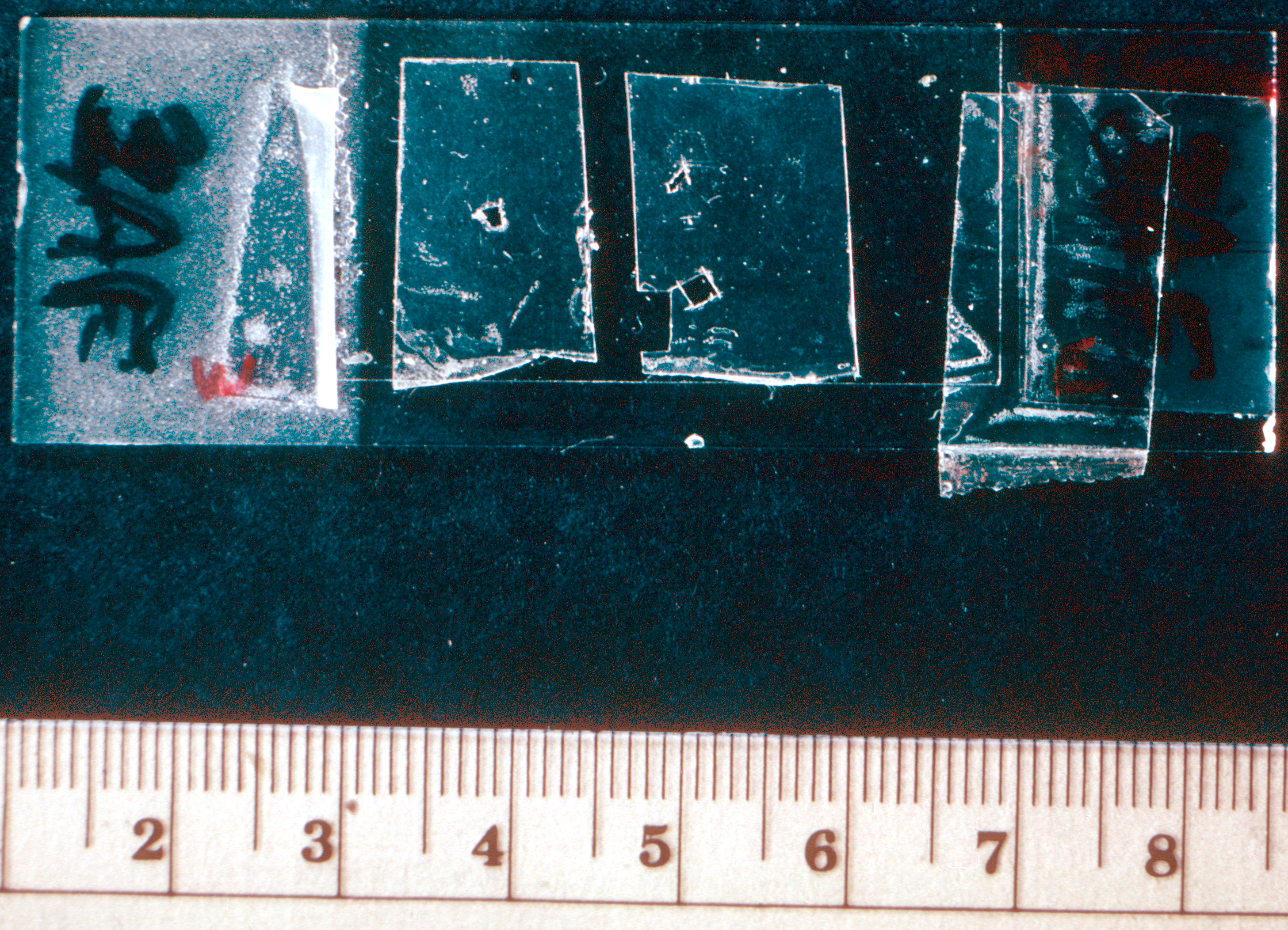
Here are a some photos of fibres from the Shroud image, from Walter McCrone and Eugenia Nitowski.

Top two: McCrone: The Shroud of Turin: Blood or Artist’s Pigment
Bottom two: Eugenia Nitowski (private collection)
A significant feature of most low power photos of Shroud slides is the debris field, as well as what appears to be particles adherent to the fibres, which is best resolved at higher magnifications. It is unfortunate that such was the acrimony between Heller and McCrone that the former was led to attempt to win the field by his misleading press release, which has dominated authenticist dogma since. In their own paper on the subject, Heller and Adler identified red particulates from submicron to 3µm in size, the smaller, to about 1µm, being identified as iron oxide. They even saturated some control cloth in 1µm iron oxide particles – 0.1% Fe2O3 suspended in “sonerated” 0.1% gelatin – and achieved: “Fe2O3 particles frequently clumped as well as adherent to fibrils, resembles birefringent red particulate Shroud fibrils more closely than the non-birefringent red coated fibrils under phase contrast; no internal particles seen in the lumen of the fibrils.”9 This is as close a description of McCrone’s own observation as to be almost indistinguishable.
A more sensible question, and one worthy of serious consideration, is whether there is enough pigment to make a visible image. The amount of iron estimated on the nose by Morris, Schwalbe and London was about 17µg/cm2, which converts to about 24µg/cm2 of red ochre.10 As investigated in a previous article, this is very easily visible on a white background. On the other hand, the same authors also estimated more than twice as much iron on an unimaged area, to the side of the “heel dribble,” which, if it were all part of red ochre, would be seen as a very obvious red patch. The iron in that area must be accounted for by some other chemical compound. When I repeated my experiments with yellow ochre instead of red, for example, I found that 24µg/cm2 was not distinguishable from unmarked cloth. In his first published paper, before his position became both entrenched and intractable, McCrone himself decided that “we now believe that the image is made up of uniformly stained fibres plus an iron oxide pigment. The latter may have been added to form the image or to enhance an earlier uniform yellow stain.”11 He later changed his mind about this more than once, but in his last paper, in Accounts of Chemical Research, says: “Microscopically the image consists of yellow fibres and red particles; the red particles are more abundant in the red blood images, and the yellow fibres are the major coloured substance in the body image.”12
From this point of view, the nature and quantity of the pigment is not really relevant to the actual image, which seems to be made more of “yellowed” fibre than of superficial granules. To this we now turn. McCrone decided it was due to a collagen tempera medium, Heller and Adler saw only dehydrated cellulose, and I think a number of alternatives are also possible, from an iron stain such as ferrous sulphate or acetate to tannins involving oak bark or galls. Medieval workshops were cornucopiae of both well tested and experimental dyes and “steyns,” and numerous recipes have come down to us.


In the absence of any thorough analysis of the image as a whole, or at least as a coloured area on a textile rather than fragments of fibres seen through a microscope, it seems futile to be too precise about the constitution, viscosity and application of whatever originally determined the image, especially allowing for the possibility that it may not now look much as it did seven hundred years ago. The best we can do is examine Mark Evans’s photos, which at least show short lengths of whole threads. Giulio Fanti, for example, studying a photo from the foot, sees the image as a composite of coloured and uncoloured fibres, the colour on any particular fibre varying in intensity along its length, sometimes ending abruptly.13

Note that the discolouration follows the fibres along the threads, which is exactly what capillary action might be expected to produce. My own analysis, of another of Evans’s photos, shows that both the colour and intensity of the image varies distinctly, if not very greatly, both along and across the threads.

Attempts to examine the fibres more closely are not necessarily more instructive. By the time Slide 3AF (from the fingers of the right hand) arrived at Eugenia Nitowski to be photographed, it looked like this:
Here are some photos of the fibres, X100:

It is difficult to tell much from these. There is a copious particle field, but no great quantity appear adhering to the fibres; and the fibres themselves do not appear uniformly discoloured.
The only other photos of “image fibres” are by Giulio Fanti. These are from Microscopic and Macroscopic Characteristics of the Shroud of Turin Image Superficiality, Slide 1EB, the back of the calf, and illustrate certain features that can be found under some conditions:

At the top (Fanti’s Figure 9.C), he points out where the Primary Cell Wall is rucked and folded over itself, so the the colour it carries appears more intense (features 1, 3 and 4), and, demonstrating that the colour is restricted to the Primary Cell Wall, as the inside of the fibre appears colourless where the PCW has been broken and stripped away (feature 2). At the bottom (Fanti’s Figure 13), he shows how the colour of any particular fibre can fade from “image colour” to “non-image colour” along the length of a fibre.
At this point I think it sensible to inquire into what is meant by “uniformity” which some people think describes the Shroud image, without, usually, being able clearly to explain what the term might mean. It has been thought, for example, that the fibres of the image were dichotomously either “coloured” or “uncoloured” and that all the coloured fibres were identical and all the uncoloured fibres were identical. The apparent variation in intensity, then, was simply a question of the proportion of coloured to uncoloured fibres in any particular area.

Another possible way of producing variation would be if all the fibres in a particular area were coloured, with the colour itself varying in intensity.

The truth, of course, as can be seen from the Mark Evans photos, is a mixture of the two, with variations along the fibres as well.
Finally, the last major claim of the miraculists is that “there is no image under the blood.” This has become such an ingrained fact that in a recent podcast Mark Guscin rather lost interest in any possibility that the image might be artificial simply because neither Luigi Garlaschelli, nor myself, thought that this fact is at all established, while he thought that no artificial explanation is possible without it.
In their Chemical Investigation paper, Heller and Adler classify the fibres they observed into seven categories, including “very pale” (backing cloth and patches), to “pale yellow” (non-image areas), “yellow” (image), and “golden yellow” (found at blood margins). I’m afraid I think these are largely unreal. Observation of any of Mark Evans’s micrographs shows that there is a continuum of colour from pale to dark along and between fibres all over the Shroud, and no evidence at all of any distinct “blood margin.”
In addition, Heller and Adler find “red coated” fibres, found in blood areas. We are perhaps to imagine either that when the blood was applied a layer of serum covered the fibres, itself covered, but not quite to the same extent, by a layer of blood, or that the blood and serum separated, so that the blood fibres were covered just in blood, while the serum alone covered the fibres surrounding them. Neither of these is evidenced by actual observation. Blood fibres, in particular, are not “red coated” at all; they are more or less uncoloured, but dotted with adherent clumps of red particles.

It is in this light that we need to read what Heller and Adler say in their Chemical Investigation paper: “One further direct and specific test was carried out for proteins namely the effect of proteolytic enzymes. For these purposes a fresh concentrated mixture of trypsin, chymotrypsin. carboxypeptidase and lysozyme in pH 8.4 buffer was employed. Within a half hour this solution completely “dissolved” the non-birefrigent red particulate coated fibril coatings, leaving no particulate residues. This further indicates that these particulates are blood and not Fe2O3 impregnated protein binder. This protease treatment also removes the golden yellow coating from the golden yellow fibrils, corroborating their identification as “‘serum” coated fibrils. Interestingly. fibrils freed of their coatings using this technique closely resemble the non-image fibrils when viewed under phase contrast. […] Proteases had absolutely no effect on the yellow (body) image or pale yellow non-image fibrils of the Shroud.”14
Adler restated this in slightly different terms twenty years later in an article at Shroud.com:
“Sticky tape non-image, image, and serum coated fibers were extracted from the tapes, cleaned, and characterized as in previous studies and tested along with a number of fibers from the radiocarbon threads employed in the FTIR studies. The protease was only active against the serum coated fibers and as in the previous study revealed smooth, non-corroded fiber surfaces indicating that the blood images went onto the cloth before the image forming process and protected the underlying cloth.”15
Against this we have the observation of Eugenia Nitowski:
“Alan Adler photographed the surface of Shroud image fibers and noted that they appeared corroded. This was not reported, to my knowledge, by anyone else, however, on my own Jerusalem Test Cloth 4, I found image fibers to be corroded, but I never observed this on Shroud fibers.“16
(Bolding mine)
In the papers he left on his death, now published as The Orphaned Manuscript, it is clear that Adler had come to a view regarding the image that is so different from what anybody can observe that he was clearly not only not impartial, but so invested in authenticity as to misremember his own observations. In the chapter called ‘The Nature of the Body Images on the Shroud of Turin,’ he says [italics mine]:
“The image only goes one fiber deep [he had no way of knowing this, and it isn’t true], lying on top of the crowns of the threads of the weaves [no; as we can see from the Mark Evans photos, the image dips deep into the interstices between the threads] (unlike the blood images which do penetrate the cloth as they are an “applied” material). The fibers are not cemented together (no binders present) [there is no reason why a thin binder such as tempera should show such cementation], but the image process shows no signs of capillarity [yes, it does; it preferentially lies along fibres, rather than across them, and flows deep into the crevices as noted above], i.e. the image does not appear under any crossing fibres [he had no way of knowing this] and the image fibres are very brittle and show “corroded” surfaces (as would be expected for dehydratively oxidised material). All the colored fibers are uniformly colored, i.e. an exposed fibre is either colored or not colored [this is not true]. This demonstrates that the image seen at the macroscopic level is an areal density image and not a pigment concentration image. Shading is not accomplished by varying the color, but by varying the number of coloured fibres per unit area at the microlevel. [This too, is clearly untrue: see below].”17

Mark Evans’s photos of the eye (left) and the foot (right)
Claiming that the coloured fibres on the right are not darker than those on the left is clearly absurd.
Anyway, abundant experiment suggests to me that the base for the stain could be iron acetate, a simple concoction made of rusty iron and vinegar, which can end up various shades of yellow-brown, and even when filtered, tends to contain suspended iron oxide particles. Soaking oak-bark (or crushed galls) in water also produces a brown stain, but combining the two results in the very black ink popular throughout the middle ages, but the wrong colour for the Shroud.18
So here we go. After all this preamble, I reach the kernel of this paper, as it were, which was a response to a request from Giulio Fanti that I actually send him some of my experiments for review. While I was not confident that they would be examined with a wholly impartial eye, I didn’t think I had anything to lose, and sent him these:

I thought it worthwhile to send some experiments printed on my rather precious Shroud facsimile cloth, so instead of using my large “apple and grapes” bas relief (above), I used a much smaller brass dragon, about 10cm x 10cm (below), as my substrate.

Both samples were prepared identically. As described to Fanti,19 “The ‘recipe’ for the tempera began with 1 egg yolk, 50cm3 water, 50cm3 malt vinegar stirred together. Then 5ml of this mixture was added to 0.5g of yellow ochre to make the ‘paint.’
The fabric was stretched over the bas relief and taped down, and a pad of folded linen was first dipped in the ‘paint,’ and then dabbed on a piece of scrap material until only a small amount was transferred, and then dabbed on the sample piece, building up the image.”
The difference between the two is as follows, “After it was dry, one sample was lightly washed to remove surface debris, and the other washed and scrubbed with a toothbrush to remove as much pigment as possible. Both samples were then ironed to remove creases.”
The first thing to note is that both my samples show a certain amount of seepage through to the back. As can be seen, the paint was applied quite thickly, and perhaps a rather lighter hand would be required to get both a more discriminatory image on the front side, and also less seepage through to the back. If there is any image on the back side of the Shroud, it is extremely indistinct.
Next, let’s have a look at the ‘negative’ and ‘3D’ manipulations.

The unscrubbed version seems reasonably good, but the scrubbed version is much less impressive. This suggests that the pigment to medium ratio was too high, or that insufficient time has elapsed for the staining effect of the medium to take effect. Again, ever the optimist, I see these as pointers to future improvement rather than total failure.
A critical factor, it seems to me, is whether the ‘image’ fibres are distinguishable from ‘non-image’ fibres even in the absence of pigment. We will come to this in due time, as we examine Giulio Fanti’s analysis of the samples I sent. His paper, published barely a month after he received the samples and three days after being “received” by the publisher, is ten pages long, of which barely two consider the samples he was sent.20 There’s a page and half listing 42 references, of which he is the sole or co-author of 22, and the first half dozen pages are devoted to explaining, in detail, why the Shroud must be authentic, and so therefore, no artificial means of production can be correct. This typifies Fanti’s position in many of his papers on the Shroud, and, in spite of his credentials as a Professor of Mechanical Engineering, it casts doubt on the probability of an impartial assessment of the evidence. However committed a scientist may be to a particular point of view, he does not champion it as infallible when he critiques a challenge to it. You can deny, but you do not easily refute a counter-argument to anything by starting with the statement that it’s wrong. Fanti’s principal attitude, as expressed here and elsewhere, is that any attempt to model Shroud image formation must be a failure because the Shroud is authentic, and as I say, he devotes well over half his paper to establish authenticity first, without considering the samples at all.
In order to try to demonstrate some of the features of my samples, I will use another little sample, this one quite heavily stained so as to make the characteristics clearer under a microscope. Here is the sample (about 6cm x 4cm), two warp threads extracted from the lower edge, half stained and half not, and one of those threads teased apart so that we can look at individual fibres from each section.

When Fanti finally gets round to it, he defines a list of 12 criteria against which the samples are to be judged, which are themselves more dependent on a conviction of authenticity than they are of observation. Here they are, with his comments in green and mine in blue.
E1. The body image has the normal tones of light and dark reversed so the body parts nearer the cloth are darker. This fact leads us to state that the body image appears as a photographic negative.
E1 (reversed tones) and E2 (3D) can be accepted.
Fanti accepts that the “negative” effect of my experiments compares adequately to the Shroud, for which I am thankful, but his statement is incorrect. On the Shroud, the nose is darker than the cheeks, and the torso varies in shade from top to bottom and from side to side. If this is a reversal of the “normal tones,” then normal people have lighter noses than they do cheeks, and the skin of their torso varies in shade from top to bottom and from side to side. But they don’t. Apart from the effects of tanning, and local epithelial discolourations, our skin is quite uniform in colour. Where there are differences in “normal tones,” such as hair and bruises being darker than skin, we find that there is no reversal on the Shroud. They are dark in life, and they are dark on the Shroud.
E2. The luminance distribution of both front and back images can be correlated to the clearances between the 3-D surface of the body and the covering cloth. This is why the TS is a 3-D image.
E1 (reversed tones) and E2 (3D) can be accepted.
Fanti grants that some 3D effect in apparent in my samples, which I think is generous, but again, the criterion is confused. It contradicts statement E1 above. Either the colour on the cloth relates to the “normal tones of light and dark” on a human body, or it relates to the distance between a part of the body and an enveloping both. It cannot do both, except where coincidentally, a normally light part of the body is also more protuberant than an adjacent part.
The hair and beard, and the bruises on the cheeks, would be darker on a real man than the skin, and are darker on the Shroud. Their relative brightness is not reversed, as it would be in a negative. They appear dark not because they were actually white, but because they were protuberant.
Furthermore, if the intensity of colour is pre-defined as related to the distance between a body and a cloth, then regardless of its shape, the position of the cloth is bound to fit it, by simple extrapolation.
E3. Image distortions of hands, calves and torso correspond to the wrapping of a man in a sheet. Therefore, considering Ref. 1, the TS wrapped a dead human body.
E3 (distortions) are not easy to determine in a bas-relief.
No, the image distortions on the Shroud do not correspond to the wrapping of a man in a sheet. Fanti has spend many hours wrapping “various statues” in semi-transparent copies of the Shroud, and invariably created wrinkles and creases which have no representation on the Shroud at all. The Ref. 1 referred to3 falls into exactly the same logical fallacy as this one, basically saying that because the Shroud is genuine it cannot be an artificial creation, and therefore any ‘body’ it may have wrapped must, without further discussion, have been a real human one.

One of Fanti’s experiments wrapping a body.
There are numerous creases and wrinkles which have no representation on the Shroud of Turin at all.
E4. Some bloodstains also appear outside the body image.
E4 (bloodstains) are intentionally forgotten to oversimplify the experiment.
It wasn’t oversimplification at all, The bloodstains are not involved in image formation.
E5. The body image has a resolution of 4.9 ±0.5mm but no well-defined contours. This means the body image seems to disappear if one looks at it from a distance closer than about one metre.
E5 (contours), even if the scale effect must be considered, shows contours that are too accentuated to infirm that’s an image similar to the TS has been reproduced.
I think by ‘contours,’ Fanti means ‘edges.’ As my experiments were on a much smaller scale than the Shroud, the resolution is in most places much better than 5mm, and the edges correspondingly more defined. My images were also darker, and thus the contrast greater than the Shroud, so you have to get much closer before distinguishing the image from the background becomes difficult.
E6. The front image, at least near parts of the head, is doubly superficial. This means that the 0.34-mm thick-fabric presents a superficial image on one side( about 0.03 mm thick), no image in the middle and another superficial image on the other side.
Regarding E6 (double superficiality), we find an effect different from that of the TS image. In some areas of the back of the linen fabric, we find some spots consistent with the image of the front side, but these spots do not appear because of a double superficiality. Instead, they appear because the colour has been absorbed in the whole thickness of the fabric, thus passing from one side to the other.
I don’t believe in “double superficiality,” and have not come across any evidence for it. We can see, and Fanti himself points out, the dark image fibres disappearing deep into the clefts between the threads, and there is no reason to suppose that, if it does emerge on the other side, the colourant does not simply seep through.
E7. The colouration does not appear under the threads where they cross in the weave of the cloth.
Evidence E7 (crossing threads) is verified in many cases.
This is, I find, variable in quite a consistent way. Where the colouration seems to continue into, and under the crossing threads, there it reappears on the other side. Where it does not manage to find a way through, then it does not reappear on the other side.
E8. The image fibres are adjacent to non-yellowed fibres: striations are evident.
Evidence E8 return (striations) is not verified except in some very rare cases that cannot be considered typical of the result analysed.
I don’t think this is true. Especially on the ‘scrubbed’ sample, some areas are clearly lighter than others, although the applied colourant is uniform. The two possible causes for the variation in brightness are described and illustrated above, in the discussion of uniformity. Either some fibres are coloured and others not, in a dichotomous way, or there is a variation in colorant across all the fibres visible in a single thread from lots to zero. The truth, as we shall see, is a mixture of the two.

Enlargement of part of the 6cm x 4cm sample illustrated above.

Two transmitted light and one reflected light image showing “striations.”
E9. Colour is frequently concentrated in the crevices where two or more threads cross each other.
Evidence E9 (crevices) fails; indeed, a position of the colour is accentuated in proximity to the protuberances of the thread with respect to the plain of the fabric.
No. As we can see, my colour dives just as deeply into the clefts between threads as does the colour of the Shroud.
E10. There is no cementation between fibres or signs of capillary flow in the image areas.
Evidence E10 (cementation), although not frequent, contrary to the TS image, is found above all in the areas of more intense colour.

Quite true. Probably too high a proportion of egg yolk, although Fanti is gracious enough to admit that cementation is not frequent. I suspect that he studied a much larger proportion of my image than he ever has of the Shroud image.
E11. The linen fibres of the image lie only on the uppermost portions of the threads, leaving the inner fibres uncoloured.
Evidence E11 (uppermost portions) is not [verified in many cases].
As I have already demonstrated, it is actually quite difficult to get paint to soak through an entire thread. It doesn’t happen on the Shroud, and it doesn’t happen in my experiments.
E12. The fibres are uniformly coloured around their cylindrical surface, whereas variations in colour intensity can be detected along the fibres. The colour is not concentrated in spots as we could expect from a pigmented fibre.
Evidence E12 (uniformly coloured) is perhaps the most important way to highlight the complete non-conformity of the proposed method with respect to the characteristics of the TS fibres, which are uniform coloured without showing pigment traces.
This is the true crux of the possible difference between the “paint” versus “radiation” hypotheses. Fanti illustrates the ‘failure’ of my experiment with the following micrograph.

And it appears he’s quite right. The colour appears to be entirely a feature of the yellow ochre particles, and not at all thanks to discolouration of the fibre itself. On the other hand, Fanti does not show what a non-image fibre looks like, so we can’t be sure if the conclusion is correct. Here, then, are increasingly large magnifications of the coloured and uncoloured fibre tangles in the sample photographed above. Note that, unlike almost every other comparison photo of Shroud fibres, whether by McCrone, Adler, Nitowski, Kohlbeck, Adler or Fanti, all these photos were taken at the same time (over a few minutes), of the same slide, using the same illumination, so that a valid comparison can be made

Firstly, even I am surprised by the apparent lack of pigment particles. There are far fewer than I was expecting, given the thickness of the colour applied, and apparently obvious from the photo of the whole sample. Consequently, we can very clearly see that the chromophore is not principally created by pigment particles.
To try to track down the elusive particles, we can close the light input aperture, which tends to increase the contrast and the depth of field, and introduces diffraction effects suggesting otherwise invisible characteristics.

Sure enough, a handful of particles have appeared, and no doubt contribute to the overall colour, but these photos bear comparison with Shroud fibres.

Compare this to one of Eugenia Nitowski’s photos:

Now, what are we to make of all this? The first conclusion I draw is that we really don’t know enough about the microscopy of the Shroud fibres to be able to make any genuine comparison, at a microscopic level, between any experiment and the Shroud. The second is that in the absence of any precise description, these experiments seem to me as close as I am likely to come.
Of Giulio Fanti’s 12 criteria, then, this is my take:
E1 – Shroud is pseudo-negative – My experiment conforms
E2 – Shroud is 3D – My experiment conforms
E3 – Shroud distortions fit actual man – Untrue or unverified
E4 – Blood – Irrelevant
E5 – Shroud image has blurred edges – My image fails due to its small scale
E6 – Double Superficiality – Untrue or unverified
E7 – No colour under threads – Untrue or unverified
E8 – Shroud has ‘striations’ – My experiment conforms
E9 – Shroud colour is found in clefts – My experiment conforms
E10 – Shroud shows no cementation – My image fails due to too much egg
E11 – Shroud image doesn’t penetrate the threads – My experiment conforms
E12 – The nature of the discolouration – Difficult to be sure…
But have I won a million pounds? David Rolfe’s criteria are more comprehensive, but as regards the image…
R1 – Colour only darkens Primary Cell Wall – My experiment conforms
R2a – All Shroud fibres are either uniformly similarly coloured or not – Untrue or unverified
R2b – The Shroud is 3D – My experiment conforms
R3 – The Shroud fibres are uniformly coloured around their circumference – Untrue or unverified
R4 – The Shroud front and back images show the same intensity – My modus operandi conforms
R5a – The Shroud has no visible pigment particles – My experiment conforms
R5b – The Shroud has no visible stain – Untrue or unverified
R6 – Blood – Irrelevant
R7 – The Shroud is pseudo-negative – My experiment conforms
R8 – The Shroud uses medieval techniques and materials – My modus operandi conforms
At the end of it all, Fanti is quite correct to conclude that I have not reproduced the Shroud image, but I don’t agree that his analysis has shown “how far this result is from the actual production of a copy of the most important Relic of Christianity.” I think it’s come quite close.
1 From the famous “d’Arcis memorandum,’ reproduced in Ulysse Chevalier, Étude critique sur l’origine du st. suaire de Lirey-Chambéry-Turin, 1900
2 Chevalier, op. cit.
3 A Summary of STuRP’s Conclusions, at shroud.com
4 Larry Schwalbe and Ray Rogers, ‘Physics and Chemistry of the Shroud of Turin,’ Analytica Chimica Acta, 1982
5 Walter McCrone, ‘The Shroud of Turin: Blood or Artist’s Pigment,’ Accounts of Chemical Research, 1990
6 “Most iron red pigments are very finely divided (less than 1 micrometre) and appear orange in colour.” ‘Red Pigments,’ Pigment ID, Peter and Ann Mactaggart, International Academic Projects, at academicprojects.co.uk/red-pigments.
7 Schwalbe and Rogers, op. cit.
8 ‘Report of Turin Commission on the Holy Shroud’
9 John Heller and Alan Adler, ‘A Chemical Investigation of the Shroud of Turin,’ Canadian Society of Forensic Sciences Journal, 1981
10 Roger Morris, Larry Schwalbe and Ronald London, ‘X-Ray Fluorescence Investigation of the Shroud of Turin,’ X-Ray Spectrometry, 1980
11 Walter McCrone, ‘Light Microscopical Study of the Turin Shroud,’ The Microscope, 1980
12 McCrone, 1990, op. cit.
13 Images from Giulio Fanti, ‘Microscopic and Macroscopic Characteristics of the Shroud of Turin Image Superficiality,’ Journal of Imaging Science and Technology, 2010, re-captioned by me.
14 Heller and Adler, op. cit.
15 Alan Adler, ‘The Nature of the Body Images on the Shroud of Turin,’ 1999, at shroud.com
16 Eugenia Nitowski (Sister Damien of the Cross, OCD), ‘Criteria for Authentication: A Procedure for the Verification of Shroud Samples,’ 1986, privately circulated
17 Alan Adler, The Orphaned Manuscript, 2002
18 The possibility of the use of oak gall ink is discussed by STuRP member Joseph Accetta in ‘Origins of a 14th Century Shroud Image.’
19 Description in letter from HF to GF, accompanying samples, 2025
20 Giulio Fanti, ‘Turin Shroud: Comprehensive Impossibility for a Work of Art,’ Medical & Clinical Case Reports Journal, 2025
As I say, I don’t think the Shroud was made as an altar-frontal as such, even though it was displayed in front of a large altar. My overall explanation is not a complete account of why it looks the way it does, but a reasonable possible explanation. Quem Quaeritis cloths did not “need” to be 4m long – but that would be a good length to hold out in front of an altar. And they didn’t “need” to have an image or even two, but that is one possible way of adding novelty to an old tradition.
My comment at (6) was based on your statement that you think the image might have formed by some natural process. If so, then possible natural processes can be envisioned and some ideas modelled. Neither Vignon nor Rogers were able to come up with anything better than what the medievalists have come up with, so at least to some people they may be said to have utterly failed. I think this attitude is a bit arrogant on the part of the miraculists, who don’t have to devise anything ‘natural’ at all, nor anything repeatable, nor anything at all scientific. I don’t think either Vignon or Rogers “utterly failed,” and suggest that anybody who thinks they were on the right track makes some attempt to improve their method. It’s a pity that they have been largely driven underground by the miraculists.
Best wishes,
Hugh
Hugh,
Thanks for your response. You definitely made some valid points. But you may also have been a little too quick. Some of your statements are questionable, and you left some matters unaddressed.
You asked me to google “medieval altar frontal cloths,” so I did. You are mostly right about the lack of such cloths with empty spaces on their sides. Several such do show up on the screen, but appear to be certainly or very possibly modern ones, accidentally included by Google Images. And sometimes it’s hard for me to tell modern from medieval at a glance. I may also have been remembering the modern altar cloths from a google search I did a year or more ago. Mea culpa.
More to the point is my second claim, which you didn’t comment on, about many medieval altar frontals being filled with “artistic clutter” (if you’ll pardon that latter term). And so they are. Most, actually. Almost all. The truly medieval ones are full of decoration, completely unlike the Turin Shroud. Floral, animal, geometrical, human, etc. And the human figures depicted are all done in a childish style, typical of medieval art. Stick figures, awkward postures.
Regarding your statement that the dorsal image can be explained by the need to fill in the sides of a 4 meter long cloth, I find that questionable. If the frontal body image itself was some 1.75 meters long, as it apparently is, that would completely fill up the central area and spill over into the side sections of the cloth. The sides would then each have had only just over a meter of empty space, not much at all in comparison with a very large and imposing 1.75 meter front body imprint in the middle. So, no big “empty” side spaces needed filling. By the way, you seem to have missed the buttocks question, the life-size buttocks question.
And if the Turin Shroud was not a true altar cloth, as you also suggest, but rather a Quem Quaeritis stage prop, why would it need to be 4 meters long, to fit a 4-meter long cathedral altar? Why not make it 3 meters, or just 2, to cover the front of the body? Or a squarish shape, as medieval European shrouds actually were, to wrap both front and back? According to your Quem scenario it was all just symbolic anyway. Precise, accurate proportions – and who knew what they really were? – were not necessary.
The link you provided to the relief sculpture “Christ in a sepulchre” actually seems to weaken the relief sculpture case. That sculpture’s proportions – hands, feet, arms, legs – are very unrealistic, either oversized or spindly. Besides, even the best of medieval paintings do not compare with the Turin Shroud image in its realism (including the bloodstains). Art historians have said so – the few who dare to comment on this taboo subject.
Also, I don’t understand your “6.) Exactly. Tell me about it. Make a tiny colored image….” comment. You seem to be alluding to Paolo di Lazzaro’s laser image formation experiments, but I don’t see what they have to do with my point 6.
It would be nice if you could provide a drawing of your “double-sided bas-relief,” or any extant depiction or documentation of a medieval version, or an actual model made by you, preferably life-size. Then dab away at it and video yourself in the process. That would help us to understand your scenario.
Your gargoyle-carving medieval bloke, moonlighting one day as an amateur shroud dabber, was certainly precocious if he created the Turin Shroud Man on his first and only try. But as the saying goes, “Beginner’s luck!”
John L.
Hi John,
1). I’ll have to see about a video of me pouncing on my little dragon! I don’t know if I can post it here or would have to put it on YouTube, but I agree that it would be a good idea.
2/3). Er, no. That’s not how a bas relief is made. Have a look at “Allysa by Sutton Betti” which you can find at https://musekits.com/blogs/muse-kits-blog/bas-relief-sculpting. This is not simply the front of a body with the back sliced off, the entire depth of the carving is only an inch or so. Also see Donatello’s Madonna and Child on the same page. Now consider a prone body of Christ in a sepulchre – something like the one at https://inventaire.patrimoines.laregion.fr/illustration/MHR91_20076602157NUCA. This is more three-dimensional than a true bas relief, but it is much flatter than a truly realistic sculpture. This one is even more so: https://www.lueneburger-heide.de/en/service/sehenswuerdigkeiten/9307/wienhausen-the-holy-sepulcher-at-cloister-wienhausen.html. It seems to me that it would be relatively easy for an artist to sculpt a figure which was life-size in the x and y axes, but maybe only 15cm thick.
4). Not an Altar Frontal in the conventional sense. The Shroud was made to be held out in front of the altar, and then draped on it, as described in the Quem Quaeritis rubrics. It may have been hung in front of the altar for a while, indeed I think it was, but that was not its primary purpose. However, when you say “Many medieval altar frontal cloths actually do have wide blank spaces on both sides of their central design,” are you sure? Try Googling ‘medieval altar frontal,’ or ‘antependium.’ There are lots to choose from, but I can’t find any with a single central design and blank space either side. Can you?
6). Exactly. Tell me about it. Make a tiny coloured smudge on a postage-sized piece of cloth with a laser, and you’ve proved beyond doubt that the Shroud is miraculous. Produce an image with pseudo-negative, 3D, superficial, etc. etc. characteristics and you’re an utter failure. I was being sightly ironic….
6.b) Seen as a combination of smudges, the Shroud is indeed covered in “telltale human mistakes.” Almost nowhere is the 3D effect consistent, but varies irregularly in intensity even across smooth areas of the body. The brow is a particularly well known bit, and there is a big hole just under one side of the bottom lip. The torso is highly irregular, as are all the limbs. You are right: it would be very difficult to maintain consistent shading by dabbing paint on, and sure enough, that’s exactly what we see.
7). We have lots of stories about them, but hardly any of their ‘laboratory notes’ as it were. These were the kind of ephemera that just wasn’t thought worth keeping, it seems, however celebrated the artist. And no, we do not have ‘dozens and dozens’ of medieval French artists, although we probably do know a few more Italian ones. The whole concept of the “artist” is a Renaissance invention: until then not even the word existed in the sense we have today. Almost all the religious artwork from the Middle Ages, with the exception of some large paintings and some small books, is wholly anonymous. To speculate that any such artist “would probably be known to some extent” is to go far beyond the evidence. Sure, some authenticists still cling to the “work of genius” using “techniques impossible even today” with an “incredible knowledge” of anatomy, physics, photography, Roman history, etc, etc, but none of it is justified. I think he was just a bloke taken off the job of carving gargoyles and misericord seats, and given the job of making the sort of imprint a dead body might leave on a shroud if any dead bodies ever did leave anything on a shroud, to make the Easter exhibition of the Quem Quaeritis shroud a bit more interesting than it was last year. Maybe he carved a new statue, but more likely he used an old one. There will be no knowing until we actually discover it, although there’s an overwhelming likelihood that it has been destroyed.
Best wishes,
Hugh
Hello again Hugh,
It seems like we’ve been down this road before, years ago, at least in some ways. But you make a few new points now, or points I missed in the past. So, back to those numbers 1-7:
1) With regard to your dabbing technique, do you happen to have a video clip demonstrating clearly what the process involves? Maybe you yourself actually going through the steps? With your little dragon bas relief, or a Ken-doll or other mini-human figure? Or best of all a life-size wooden mannequin? You say the resolution of your dragon imprint is greater than on the Turin Shroud, but that seems questionable to me. And does it really matter?
2/3) You say you envision a “double-sided bas relief” figure of Jesus. Could you be more specific? To me, at the moment, that sounds like a reclining statue of Jesus in which the top/front portion of his whole body length is intact, also the bottom/back portion intact, but with several inches missing throughout the middle plane. Is that right? That would be one very lean Jesus. Could you show us an illustration to make it clear? A drawing? Such a creation would surely have been a first in medieval art, surpassing even the traditional slender Jesus statues. Or do you know of any extant double bas reliefs?
You also envision your bas relief “body … laid on its back, and the front image made, and then turned over, and the back image made.” But if so, the artisan/s somehow missed getting complete imprints of the front of the feet, which seems odd, especially because he/they started there on the front and also had lots of time to plan and prepare his/their creation, and space to do it in.
4) As for your altar frontal cloth scenario, I recall your suggesting it a few years ago in another online forum. I forget whether I noted any problems with it then, so here are some now: a) Many medieval altar frontal cloths actually do have wide blank spaces on both sides of their central design, whatever that design may be, so, such unoccupied side spaces seem no reason to imagine a necessary imprint of Jesus’ back in addition to a frontal imprint; b) Many other medieval altar frontal cloths are cluttered with artistic detail, of which the Turin Shroud remarkably has none; c) The very few medieval altar frontal cloths showing Jesus lying dead that I have seen also show him horizontally in profile, not floating sideways, face outward, as he appears on the Turin Shroud when it is raised up horizontally and hung down vertically. That perspective would seem to be unique if it really is an altar cloth.
Then there’s the “stained sheet … abandoned in the tomb” that you suggest. It’s not found in the Gospels, so it seems unlikely that a Christian artisan would have thought of it, or not a back image in addition to a front image. And surely not a life-sized naked back image with full buttocks.
5) You say that “most [Shroud] authenticists believe at least two, and often all six” positions of body and cloth. Such multiplicity seems doubtful to me, as far as I follow you. Maybe a poll could be taken at a future TS conference to get better statistics on the various views. But yes, you were responding to Giulio Fanti’s wrapping scenario, so your earlier answer was understandably limited.
6) It seems premature to label the natural image formation theory a “failure.” Several natural occurring means of getting approximations of the TS image have been found over the decades. Besides, not nearly enough experiments have been done in that regard. Few people in the field take the natural image position, so they are very limited in funds and in potential collaborators for experiments. I’ve repeatedly advocated for such experiments, but have gotten no response. Another point: The natural image theory would require only one cause, one act (with maybe 2-3 contributing factors), whereas your dabbing craftsmen scenario would require at least several hundred careful little individual dabs and over a lengthy period of time, each of them capable of going astray, of being slightly out of alignment, thus revealing telltale human mistakes to us today. Yet no such mistakes are evident on the Shroud. Not one.
7) You mention Leonardo and Michelangelo as almost without written sources on their lives, but actually there is quite a lot of contemporaneous writing extant about both, not to mention all the known artworks of each one. And there are dozens of 13th and 14th century French artists whose names are known today along with some details about them. Most people in the Shroud field would honestly say that if the Shroud is a medieval creation it was at least the work of an artist, in fact a genius. Such an artist would probably be known to some extent. Your suggestion that merely “some obscure monastic craftsmen” created the image seems itself crafted to excuse the complete absence of any known detail about them. No name, no location, no date, no payment, no request for funds, no recorded technique, no wife or wives, no children, no forebears, no abbot, no teacher, no school of students to carry on, no rivals, no nothing. That situation, that void, still seems extremely odd and suspect to many of us. But at least you write of “some … craftsmen,” in the plural. That’s realistic: a group, a team. What did Bishop d’Arcis claim in 1389? A single “artist” who apparently created it all on his own. Now that would be a miracle.
But d’Arcis deserves sympathy, too, because the clerics of Lirey were apparently using the Shroud in on-the-spot miraculous healing scams, which did not reflect well on the Shroud itself. “Guilt by association” – a common but flawed judgment.
Keep on trying, Hugh, but more carefully, please. I will too.
John L.
Hi John,
Thanks for your thoughtful and considered comments, as usual.
1). I don’t know of any medieval artwork performed in the same way as I have proposed or the Shroud. However, the concept of “pouncing” was common, but in a different context. Fresco designs were usually sketched out on paper, and then all the lines pricked with little holes. The paper was then taped to the wall, and then a small bag of red ochre, about the size and shape of those little bags of herbs cooks sometimes drop into stews for flavour, was dabbed all over it. The paper design ended up covered in ochre, and was thrown away, although it may have looked a little like the Shroud; and the design appeared on the wall as lines made of little dots.
The size of a finger is not strictly relevant to the resolution of the image, as what makes contact with the cloth is a soft pad, which in my case is usually made of many thicknesses of cloth taped to a piece of dowel, or a strip of cloth rolled into a stick about a centimetre in diameter, with one end deliberately frayed. As you can see, the resolution of my dragon images is much greater than anything we see on the Shroud. If you dip the ‘pounce’ into the colourant, and then dab it several times on a piece of scrap till it hardly makes a mark, you can then apply it to your draped linen, and dab away as much or as little as necessary, building up colour if you want. There’s no bother about sneezing and jerking, all you are doing is dabbing, rather randomly.
2/3). I don’t think I’ve ever seen a picture of the underside of any of the wooden figures of Christ that fit into the Easter Sepulchres that I’ve often mentioned in the past, so I can’t say if they are also carved, but I envisage the figure used for the Shroud to be a double-sided bas relief, so the question of whether the proportions are the same front and back is minimised. There are several prone statues of Christ extant, of which the wooden ones appear to be carved both top and bottom. I think the body was laid on its back, and the front image made, and then turned over, and the back image made.
4). I’ve wondered about that too. If the Shroud were made for display in front of a high altar, then perhaps it was just an idea to fill it to the ends, rather than just have the one image in the middle and wide spaces either side. If the Shroud was meant to display a miracle, then a single image, beautifully painted, would be appropriate, but as I have said before, I don’t think it was. It think it was meant to represent the stained sheet that might have been abandoned in the tomb after the miracle had happened.
5). Quite so. Most authenticist believe in at least two, and often all six, of six mutually incompatible designs (body flat or bent X shroud flat, draped or wrapped). My comment was specifically in response to Giulio’s response to my experiment rather than generally about all authenticists. There are some sensible ones, I suppose…
6). Yes. All power to the ‘nature’ people, and I sympathise with their failure to come up with any better way by which the image might have occurred than mine. It’s easy for the miraculists, as all problems can be swept under the “it’s a miracle” carpet.
7) Sadly, there is vanishingly little that remains of any of the ephemera of the great cathedrals of the past. We don’t who built them or who decorated them, and have vanishingly little paperwork. We have almost nothing of Michelangelo and Leonardo da Vinci, let alone some obscure monastic craftsmen.
Thanks for writing though, and keep a look-out for Dale’s next couple of podcasts, and no doubt my responses to them here.
Best wishes,
Hugh
Hi Hugh,
A very interesting post, your “How was it done?” Thanks for making the effort.
The post is mostly over my non-scientific head and non-microscopic eyes, but very few other readers have commented on it, and the Comments box is due to close in just days. So I might just ask a few questions, since I have doubts about your medieval shroud hypothesis:
1) Is there any documentary trace of your “dabbing” artistic technique (pp. 1-2, 13) in the Middle Ages up through the 14th century? Does your dabbing technique envision using a finger to press on the cloth? If so, would a finger tip, about 1 cm wide, have been able to produce the smaller, finer features seen on the body image? Time after time for hours on end, never veering off track? What if the artist sneezed? What if his black cat suddenly jumped onto the table, startling him, causing his finger to jerk, and thus marring his image? “Back to the drawing/dabbing board” after days or weeks of effort?
2) You say a bas relief sculpture was involved (pp. 1, 13, etc.). But don’t you actually mean two separate bas reliefs, one for the front and one for the back? If so, that would be a rather awkward way to go about producing the double image seen on the Turin Shroud, don’t you think?
3) I’ve read that the two images on the Turin Shroud correspond to each other perfectly, that is, the front image matches the back image exactly in all its positions and anatomical measurements – of the head, the trunk, the legs, the feet. So much so that many or most authenticity-believing scientists state that a single, complete body (let’s leave out for now whether human or statue) really must have been the object of the image formation. What is your response to that? Or did the artist first create one reclining full statue and then, in order to get two bas reliefs, take a handsaw to it, cutting it in two from head to foot?
4) What purpose would also having a dorsal image have served a medieval artist? It seems superfluous to me. A statue is, by definition, in the round, but a sculpture is not always in the round, some sculptures being mere bas reliefs, and a two-dimensional representation on wood, cloth, or canvas is certainly not in the round. So, why add the back image to the Turin Shroud? I’m thinking now also of my article, “The Back of Jesus in Medieval Painting Compared with the Turin Shroud,” in the Summer 2023 issue of the BSTS Newsletter. It was far from exhaustive, but I looked randomly at several hundred medieval illustrations of Jesus and found only a single one that depicted his full back or dorsal aspect. It was tiny too, his figure being only about 10-12 cm. high, and was not at all realistic in its body proportions (the 9th century Stuttgart Psalter scourging scene). As I speculated, there may actually be one or two illustrated “backs of Jesus” out there in some obscure medieval manuscript or church closet, but even if so, they would presumably be on the order of one in 10,000 medieval pictures of Jesus, virtually unique. And they would also probably be tiny or small in size. So, why would reproducing the full back view, and in life size, even have been conceived by such a medieval artist as yours?
5) You show and critique Fanti’s simulations of a tight shroud body-wrapping (p. 15), noting that no such major distortions are evident on the Shroud image. Well done. But that’s Fanti’s scenario. Others have long proposed a fairly flattened top sheet propped up by sacks of spices or other such materials, which fortuitously permitted a relatively undistorted frontal image.
6) On page 10 you write of the “miraculists” as your opponents or discussion partners in the debate, but some of us others do not believe the image was formed by a miracle, only Nature.
7) How many years have you now spent in theorizing and attempting to reproduce such a mere “general characteristics” copy of the Turin Shroud? I’m thinking you’ve been at it day and night for about ten years. That’s a long time and a lot of energy. You must have records of your efforts too. Surely any medieval artist or craftsman would also have taken years, would have kept some records, would have spoken with fellow artists of his creation-in-the-works, yet no such records are extant, no reliable testimony, no reliable witness. And did the artist finish his masterpiece and then simply keel over dead, never going on to produce any other such powder-puff marvels, similar versions of, say, the apostles, the Mary’s, the saints? Surely the clerics would have been eager for him to do so. Yet, there’s no reliable trace of him in recorded history. Hmmm.
But your attempts are certainly laudable, Hugh, and you definitely poke holes in some lazy authenticist claims. Thanks for that.
John L.
Quick comment on painting. I’m an amateur painter. I bought a book many years ago on how to paint like the masters. One of the things mentioned was the importance of priming one’s canvas with rabbit skin glue and then with lead white paint. Anything less is pretty much going to be inferior. The book mentioned how the typical canvases that one purchases at the art supply store are total junk, because the don’t properly prime the canvases (whether cotton canvas or linen.) It’s very difficult to find canvases primed with lead white (also known as “flake white” paint. Usually, the white paint used on canvases in titanium white. Anyhow, poorly primed canvases result in even oil paint (far more viscous than watercolor paint) seeping into the canvas and not sitting “on top” of the canvas the way that a properly primed canvas will allow for paint to lay upon it.
Here’s the show where Hugh and I discuss his newest attempt at unraveling how the body image was formed. https://youtu.be/RRwIf229ZEc?si=QRIep0YJm8kYyGdo
Hugh and I got into the weeds with the London, Morris and Schwalbe paper. I go into the details of this paper in the other show that I have linked to in my prior email (the one with Jack Markwardt and Dale.)
Hi, Hugh and Richard,
I gave a brief response on Dan Porter’s Shroud website that references this posting of yours, Hugh. Also, the show that you and I did right before Christmas (on Dale’s podcast Real Seekers Ministries) has a lot of my responses. Additionally, more of my responses can be found on the show Jack and I did on Dale’s podcast.
I’m overly swamped with not one but two papers (that I have no business taking on since I’ve still got a book to finish writing), so I have not had a chance to read your posting, but I figure it’s about what we discussed on Dale’s show right before Christmas.
So, for people who would like to examine what some of the counterpoints are, I give this link and I’ll post the other one in a separate post.
Cheers,
Teddi
Here’s one of the links. https://www.youtube.com/live/tIfi2TyjQB8?si=d5-UyxWTEuURDYBR
Very true, although it’s easy to arrange one’s materials so that paint doesn’t seep through whether the canvas is primed or not. Did anyone at
STuRP experiment to find out, I wonder?
great work, as usual, Hugh. I feel I should point out though that the reason that paint doesn’t normally penetrate through to the back of its canvas is that the canvas will have been primed, usually with rabbit skin glue, which provides a fairly robust barrier to paint. as far as I know, there is no such priming on the Shroud?